
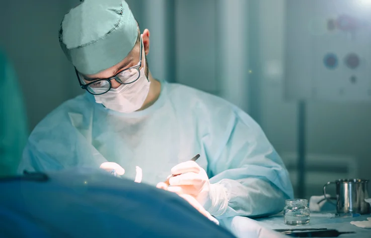
In the high-stakes environment of the modern operating room, the margin for error is razor-thin, and the quality of real-time information can dictate the trajectory of patient outcomes. Traditionally, surgeons have relied on two-dimensional scans—standard X-rays, Computed Tomography (CT), and Magnetic Resonance Imaging (MRI)—to visualize three-dimensional human anatomy. However, a technological paradigm shift is currently underway. At leading medical institutions, surgeons are no longer limited to mental reconstructions of flat images; instead, they are utilizing sterilized 3D-printed heart models, digital airway reconstructions, and virtual reality (VR) environments to navigate the complexities of the human body before the first incision is ever made.
This integration of immersive media and additive manufacturing represents the culmination of nearly three decades of innovation. What began as a niche application in dental and maxillofacial surgery has evolved into a comprehensive clinical toolset that spans neurosurgery, cardiology, orthopedics, and pediatrics. As these technologies become more accessible, they are fundamentally altering the workflow of surgical preparation, intraoperative decision-making, and patient communication.
The Evolution of 3D Technology in Clinical Practice
The journey of 3D printing in healthcare began approximately 30 years ago, following the commercialization of stereolithography in the late 1980s. Early applications were primarily focused on creating anatomical models for complex reconstructive surgeries, particularly in cases involving the skull and jaw. These early models allowed surgeons to visualize bone deformities in a physical space, providing a tactile advantage that 2D films could not replicate.
Over the last decade, the field has transitioned from basic physical models to "immersive media," a term that encompasses 3D printing, Virtual Reality (VR), and Augmented Reality (AR). The timeline of this evolution highlights a steady march toward precision:
- 1990s: Introduction of 3D modeling for dental implants and complex maxillofacial reconstructions.
- 2000s: The rise of patient-specific instrumentation (PSI), where 3D printing was used to create surgical guides tailored to a patient’s unique bone structure.
- 2010s: Integration of VR and AR, allowing surgeons to "rehearse" surgeries in a digital twin of the patient’s anatomy.
- 2020s: The normalization of in-house 3D printing labs within major hospital systems, facilitating rapid turnaround for emergency procedures.
Today, the technology is no longer an experimental luxury but a vital component of the surgical pipeline, especially in pediatric facilities where unconventional anatomy is the norm rather than the exception.
From Pixels to Plastic: The Technical Workflow
The creation of a 3D medical model is a meticulous process that bridges the gap between radiology and engineering. It begins with high-resolution medical imaging. For a model to be clinically useful, the underlying CT or MRI data must be captured with specific protocols that ensure thin "slices" of data, providing the detail necessary for accurate reconstruction.
Once the images are acquired, the process of "segmentation" begins. In this stage, 3D modeling specialists use sophisticated software to isolate specific tissues—such as a tumor, a blood vessel, or a heart valve—from the surrounding anatomy. This digital "tracing" allows for the creation of a 3D mesh. This mesh can then be refined through "reconstruction," turning a cloud of digital data into a watertight computer model.
For a digital rendering, this model can be uploaded into a VR headset, allowing a neurosurgeon to virtually "walk through" the blood vessels of a patient’s brain to identify the safest path for an aneurysm clip. For physical applications, the model is sent to a 3D printer. Depending on the complexity, a specialist can produce a computer model within minutes, while a physical, biocompatible 3D print can often be delivered to the surgical team within 24 hours.
Enhancing Precision through Patient-Specific Instrumentation
One of the most significant advancements in this field is the development of patient-specific guides and templates. Beyond simply looking at a model, surgeons can now use 3D printing to create tools that only fit one specific patient.
For example, in orthopedic surgery, a 3D-printed cutting guide can be designed to snap onto a patient’s femur at a precise angle. These guides often feature depth markers and pre-defined channels for drills or saws. By using these tools, surgeons can ensure that their cuts are accurate to within a fraction of a millimeter.
Furthermore, this technology allows for the "pre-bending" of surgical hardware. In complex spinal or reconstructive surgeries, metal plates often need to be contoured to fit the patient’s bone. Traditionally, this was done during the operation while the patient was under anesthesia, a process that could take significant time. With a 3D-printed model of the patient’s bone available pre-operatively, the surgeon can bend and sterilize the plates before the surgery begins. This reduces the need for "on-the-fly" alterations and significantly lowers the time the patient spends in the operating room.
Quantifying the Impact: Efficiency and Economic Benefits
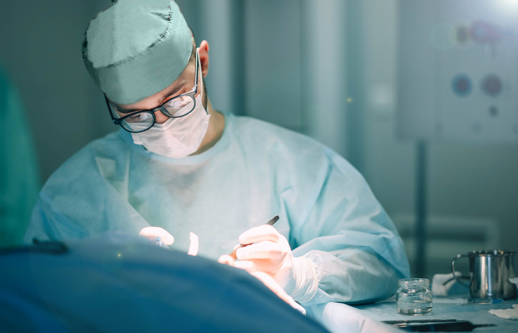
The clinical benefits of 3D modeling are increasingly supported by empirical data. Studies have shown that the use of 3D-printed models and VR rehearsals can shorten complex operations by 30 to 90 minutes. In a high-cost environment like a hospital, where every minute in the operating room can cost between $60 and $100, these time savings translate into significant financial efficiency.
Beyond direct cost savings, the reduction in operative time has a direct correlation with patient safety. Shorter surgeries mean:
- Reduced Anesthesia Exposure: Less time under general anesthesia lowers the risk of post-operative cognitive dysfunction and respiratory complications.
- Lower Infection Risk: Reduced "open-time" for surgical wounds decreases the window for pathogens to enter the body.
- Decreased Blood Loss: Faster procedures generally result in less intraoperative hemorrhaging.
A fact-based analysis of these implications suggests that while the initial investment in 3D printing infrastructure is high, the long-term ROI is found in the mitigation of surgical complications and the optimization of hospital resources.
Bridging the Communication Gap
The utility of 3D modeling extends beyond the sterile field of the operating room and into the consultation suite. One of the most challenging aspects of modern medicine is informed consent—ensuring that a patient or their family truly understands the risks and nature of a procedure.
Standard medical jargon and 2D scans can be alienating and confusing for laypeople. A 3D model acts as a "universal language." For instance, in pediatric cardiology, explaining a complex septal defect to parents using a computer screen is difficult. However, handing those parents a color-coded, 3D-printed model of their child’s heart allows them to see and feel the defect. This tactile interaction fosters a deeper level of trust and understanding, enabling families to make more informed decisions regarding care.
The Institutional Shift: The Case for In-House Labs
While 3D printing services can be outsourced to third-party medical device companies, there is a growing movement toward establishing in-house 3D printing service lines within hospitals. The "enthusiast" model—where a single physician uses a personal 3D printer for their own cases—is being replaced by integrated hospital departments.
In-house labs, such as the 3D Innovations (3DI) Lab at Rady Children’s Health in San Diego, provide a centralized resource that serves multiple departments, from oncology to orthopedics. This centralized model allows for:
- Collaboration: 3D specialists work side-by-side with physicians to tailor models to specific surgical preferences.
- Standardization: Hospitals can implement rigorous quality assurance protocols, ensuring that every model meets medical-grade standards.
- Innovation: Having the technology on-site encourages physicians to experiment with new ways to solve "unsolvable" cases.
Organizations like the Radiological Society of North America (RSNA) and the DICOM (Digital Imaging and Communications in Medicine) Working Group 17 are currently working to modernize standards to support these in-house efforts. By creating guidelines for clinical appropriateness and quality assurance, these bodies are ensuring that 3D modeling moves from an "art form" to a standardized medical practice.
Future Outlook and Systematic Integration
The future of 3D modeling in healthcare is not merely tied to better printers or faster software, but to the seamless integration of these tools into the standard medical workflow. We are moving toward a future where "digital twins"—complete virtual replicas of a patient’s physiology—will be used to simulate not just surgeries, but the effects of drugs and other interventions.
However, challenges remain. Regulatory hurdles regarding the classification of 3D-printed "devices" and the lack of universal reimbursement codes for 3D modeling services can slow adoption. Furthermore, the success of these programs depends on the willingness of healthcare systems to adapt to new processes.
In conclusion, 3D modeling and immersive media have transitioned from futuristic concepts to essential surgical assets. By providing surgeons with the ability to see the "unseen" and practice the "unpracticed," these technologies are reducing risks, saving costs, and, most importantly, improving the lives of patients. The revolution in the operating room is no longer about the tools in the surgeon’s hand, but the depth of the information guiding them. Through integrated teams and well-defined systems, the medical community is setting a new standard for surgical precision in the 21st century.

